
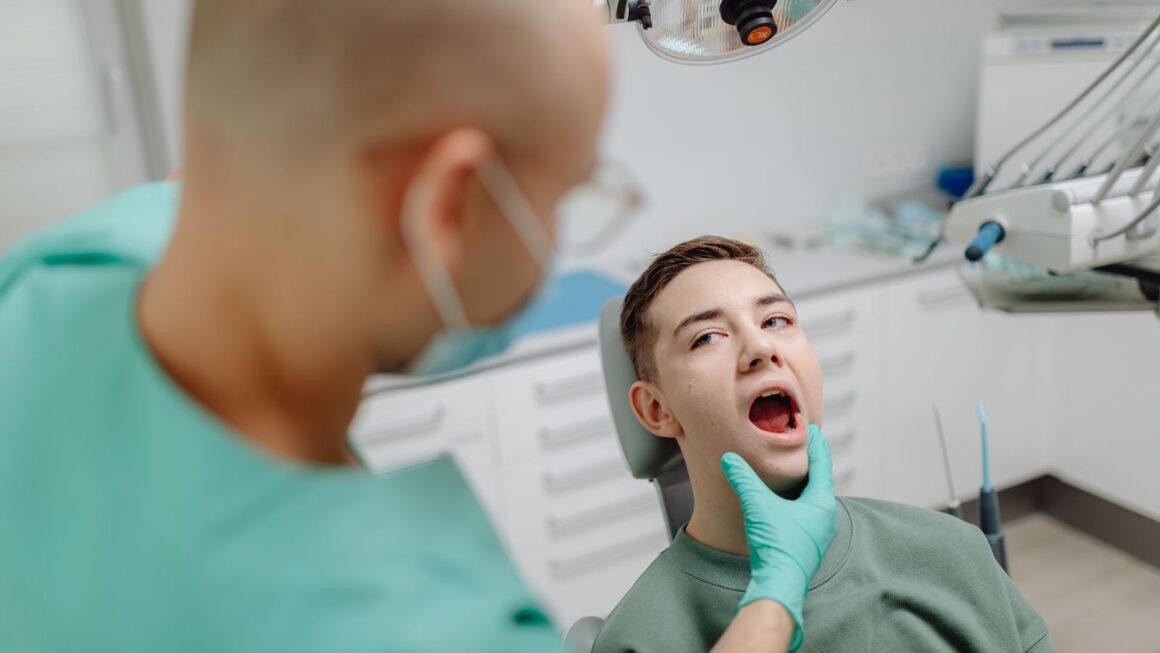
Losing a tooth starts a chain reaction in your jaw: the bone that once supported the root no longer receives stimulation and gradually resorbs. Consulting an implant dentist in Blaine, WA is one of the most effective ways to address this — bone loss from a missing tooth does not reverse on its own, and without intervention the jawbone will continue to shrink, though modern dental treatments can halt progression and often rebuild lost bone.
You’ll explore what causes that resorption, how quickly it can happen, what the science shows about natural recovery versus clinical regeneration, and which treatments and preventive steps give you the best chance to preserve or restore your jawbone.
Understanding Bone Loss from Missing Teeth
Missing a tooth removes the mechanical stimulus the jawbone needs, which leads to gradual shrinkage in the area of the gap. You’ll see changes in bone height, width, and density that affect future restorations and facial support.
Causes of Jawbone Deterioration
When a tooth root is gone, the local bone loses the forces from chewing that normally stimulate bone maintenance. This process, called disuse atrophy or resorption, triggers osteoclast activity (bone breakdown) outpacing osteoblast activity (bone formation).
Infection and periodontal disease accelerate bone loss by destroying the supporting structures around adjacent teeth. Systemic conditions such as osteoporosis, uncontrolled diabetes, and long-term smoking further weaken bone quality and slow healing after extraction.
Common causes you should watch for:
- Tooth extraction without immediate replacement
- Chronic periodontal infection at or near the extraction site
- Trauma that damages the alveolar ridge
- Systemic bone-loss conditions or medications (e.g., corticosteroids)
Timeline of Bone Loss After Tooth Loss
Bone resorption begins quickly. Within weeks to months after extraction, you can lose measurable ridge width and height at the site of the missing tooth. Most rapid changes occur in the first 3–6 months.
By 6–12 months, significant ridge collapse often becomes clinically apparent, complicating implant placement without grafting. Long-term, continued slow resorption can progress for years, altering facial contours and making dentures less stable. Immediate socket preservation or early implant placement reduces the rate and magnitude of this timeline.
Key Factors Influencing Bone Resorption
The amount and speed of bone loss depend on local, systemic, and behavioral factors. Locally, whether you had an infected socket, the thickness of your buccal bone, and the number of adjacent teeth missing matter most.
Systemic factors include age, hormonal status, bone metabolism disorders, and medications that affect bone turnover. Behavioral factors—poor oral hygiene, smoking, and low dietary calcium or vitamin D—worsen resorption and impair healing.
Practical considerations:
- Thin buccal plate → greater collapse risk
- Multiple adjacent missing teeth → larger volume loss
- Smoking and uncontrolled diabetes → slower healing, more resorption
Can Bone Loss Reverse on Its Own?
Bone loss after a tooth is lost rarely restores fully without intervention. Small, early changes can stabilize, but significant regrowth of alveolar bone typically does not occur spontaneously.
Current Scientific Consensus
Researchers and dental specialists agree that natural, complete reversal of bone loss following tooth extraction is uncommon. After a tooth is removed, the alveolar ridge undergoes predictable remodeling: most resorption happens within the first 3–6 months and continues at a slower rate afterward. Clinical studies show that without targeted treatment—such as socket preservation, bone grafting, or implant placement—ridge height and width decrease measurably.
You should expect modest stabilization if inflammation is controlled and loading patterns are restored, but true regeneration of lost volume usually requires surgical or biological therapies. Evidence supports interventions that either preserve bone immediately (grafts, membranes) or stimulate new bone formation when reconstruction is needed.
Natural Healing Limitations
Your body initiates a healing response after extraction, forming a blood clot and then woven bone within weeks. That initial bone is structurally different and often remodels into a thinner ridge rather than restoring the original shape or density.
Key limitations:
- Lack of mechanical stimulation where the tooth used to be reduces bone maintenance signals.
- Normal age-related bone turnover and systemic factors (e.g., osteoporosis) limit regenerative capacity.
- Soft-tissue collapse into the socket can prevent space needed for bone to reform.
These constraints mean natural healing preserves some bone but seldom restores pre-extraction contours sufficient for ideal prosthetic outcomes without augmentation.
Variables Affecting Bone Regeneration
Several specific factors change how much bone you might retain or regenerate:
- Timing of intervention: Immediate socket grafting reduces resorption compared with waiting months.
- Local infection or periodontal disease: Ongoing inflammation accelerates loss and impairs healing.
- Systemic health: Diabetes control, smoking status, and osteoporosis medication affect bone metabolism.
- Mechanical loading: Placing an implant or maintaining occlusal forces helps preserve bone through functional stimulation.
- Graft and biologic materials: Types of bone grafts, membranes, and growth factors influence the volume and quality of new bone.
Assessing these variables with your dental professional lets you choose predictable options—such as grafting at extraction, guided bone regeneration, or implant-supported restorations—rather than relying on spontaneous recovery.
Scientific Approaches to Restoring Lost Jawbone
You’ll find mechanical restoration, biologic regeneration, and implant-driven preservation as the primary strategies. Each approach targets different degrees of bone loss and has distinct timelines, materials, and success rates you should weigh.
Bone Grafting Techniques
Bone grafting replaces missing bone volume to rebuild ridge height and width for function and implant support. Autografts use your own bone (often from the chin or hip) and offer the best osteogenic potential but require a second surgical site and longer healing. Allografts (donor human bone) and xenografts (bovine-derived) avoid a harvest site and provide a scaffold for new bone growth; they integrate more slowly but are widely used.
You’ll see block grafts for large defects and particulate grafts for socket preservation after extraction. Surgeons often pair grafts with barrier membranes (guided bone regeneration) to prevent soft tissue ingrowth and improve graft stability. Expect 3–9 months of healing before implant placement, depending on graft type and clinical factors.
Role of Dental Implants in Preservation
Dental implants substitute tooth roots and deliver mechanical stimulation to maintain alveolar bone. When you place an implant into a healed or grafted site, the implant transfers functional loads during chewing to the surrounding bone, which slows or halts resorption through normal bone remodeling.
Immediate implant placement at extraction can preserve ridge morphology but depends on infection control and initial stability. Delayed placement may require prior grafting if resorption occurred. Success rates exceed 90% in healthy patients, but smoking, uncontrolled diabetes, and poor oral hygiene reduce predictability. Proper implant size and positioning also influence long-term bone maintenance.
Emerging Regenerative Treatments
Tissue engineering focuses on biologics and scaffolds to stimulate in situ bone formation rather than just filling defects. You may encounter growth factors such as BMPs (bone morphogenetic proteins) and platelet-derived preparations (PRP, PRF) used with scaffolds to accelerate and enhance bone regeneration.
Stem cell therapies using mesenchymal stem cells from bone marrow or adipose tissue show promise in clinical trials for larger defects, though protocols and regulatory approvals vary by region. Synthetic biomaterials with controlled resorption rates and 3D-printed patient-specific scaffolds help match complex defect geometries. These methods can reduce the need for donor bone, but cost, access, and long-term outcome data remain evolving.
Preventing Bone Loss Following Tooth Extraction
You can limit bone resorption by acting at the extraction appointment and within the first weeks afterward. Focus on procedures that preserve the socket and on timely tooth replacement to keep the jawbone stimulated.
Immediate Post-Extraction Strategies
Place a socket graft at the time of extraction when possible. Your dentist or oral surgeon will pack bone graft material (autograft, allograft, xenograft, or synthetic) into the empty socket to preserve ridge height and width. This reduces early collapse of the bone that normally begins within weeks.
Ask about a collagen membrane or primary closure if you have thin buccal bone. These measures stabilize the graft and protect against soft-tissue ingrowth. Follow post-op instructions precisely: avoid suction, smoking, and hard foods for 7–14 days to prevent dislodging the graft.
Consider immediate implant placement when local anatomy and infection status allow. A properly placed implant can provide mechanical stimulation to the alveolar bone and often reduces the need for later extensive grafting. Your clinician will assess socket integrity, occlusion, and systemic health first.
Importance of Timely Intervention
Replace the missing tooth within weeks to months rather than years to maintain bone volume. Delaying replacement increases the likelihood of ridge atrophy that complicates future implant placement or requires larger grafts.
Choose replacements that restore root function. Dental implants act like roots and transmit functional load to bone, which helps maintain density. Fixed bridges or well-fitted removable dentures can limit bone loss compared with no replacement, but they don’t stimulate bone as effectively as implants.
Monitor healing with follow-up imaging. Your dentist should take periapical radiographs or CBCT scans at appropriate intervals to confirm graft integration or bone stability. If you have risk factors—smoking, diabetes, or periodontal disease—manage them aggressively to improve outcomes.