
If your jaw lacks the height or density to hold an implant, a dental bone graft restores the support your new tooth needs so the implant can last.
A bone graft builds or augments jawbone using natural or synthetic material to create a stable foundation for dental implants, making implants possible when bone loss would otherwise prevent them.
You’ll learn when a graft becomes necessary, what the procedure involves, and how recovery typically unfolds so you can weigh risks, timing, and expected outcomes. Clear explanations of techniques like ridge augmentation and sinus lifts will help you understand options and what to expect before, during, and after surgery, especially when exploring full arch dental implants pittsburgh as a long-term restorative solution.
Understanding Bone Grafting for Dental Implants
Bone grafting rebuilds or adds bone where your jaw has lost volume or density, and it creates a stable foundation for implants. You’ll learn what grafting does, why dentists recommend it for implants, and the common graft materials and techniques used in practice.
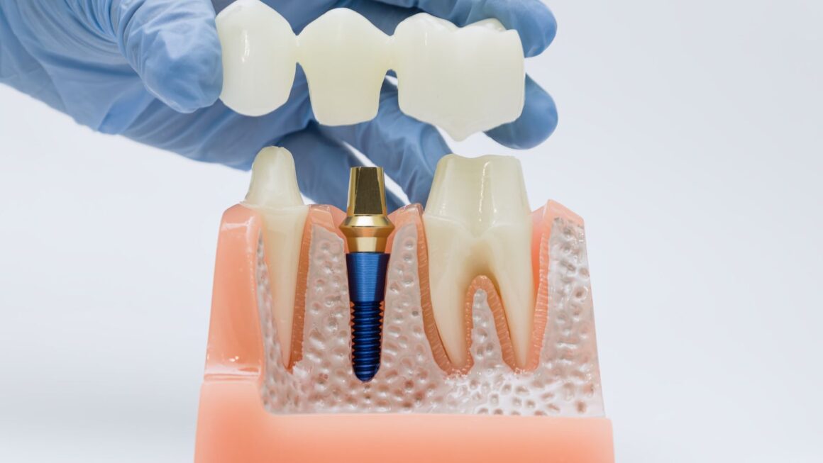
What Is Bone Grafting?
A bone graft places bone material into an area of your jaw that lacks sufficient height, width, or density. The graft acts as a scaffold for new bone growth, a process called osseointegration, which allows your natural bone to form around and incorporate the graft material.
Clinicians perform the procedure under local anesthesia, often with sedation for comfort. They evaluate your jaw with X‑rays or a cone beam CT scan to measure bone volume and plan the graft precisely.
Healing time varies by graft type and location. Small grafts can integrate in a few months; larger augmentations, such as ridge expansions or sinus lifts, may require four to nine months before implants can be placed.
Why Bone Grafting Is Required for Implants
Dental implants need at least a certain amount of bone to anchor and withstand chewing forces. If your jawbone is too thin, too short, or has been resorbed after tooth loss, the implant may fail or be unstable.
Specific situations that commonly require grafting:
- Long-term tooth loss with bone resorption.
- Periodontal disease that destroyed supporting bone.
- Trauma or infection that removed bone volume.
- Sinus pneumatization in the upper molar area reducing vertical height.
Your provider will measure bone height, width, and density. If measurements fall below implant manufacturer or clinician thresholds, they’ll recommend grafting options to achieve predictable, long-term implant stability.
Types of Bone Grafts Used in Dentistry
Dentists use four main graft categories: autograft, allograft, xenograft, and synthetic grafts. Each has different benefits for integration, availability, and healing time.
- Autograft: Bone taken from your own body (chin, ramus, or hip). It offers live cells and growth factors and has high success rates, but requires a second surgical site.
- Allograft: Processed human donor bone. It avoids a donor-site operation and acts as an effective scaffold; integration relies on your body’s remodeling.
- Xenograft: Sterilized animal-derived bone (commonly bovine). It maintains volume well and is often used where longer scaffold persistence is helpful.
- Synthetic grafts: Made from materials like calcium phosphate or bioactive glass. They avoid biological risks and can be tailored for resorption rates.
Clinicians may combine graft materials and use barrier membranes or growth factors (e.g., BMP, PRF) to enhance healing. Choice depends on defect size, location, your medical history, and whether you prefer to avoid donor-site surgery.
When Bone Grafting Is Needed
You need bone grafting when your jaw lacks the volume or density to support an implant or to restore oral health after disease or trauma. The following subsections explain common causes of bone loss, how clinicians determine if you need grafting, and when grafting is timed relative to implant placement.
Causes of Jawbone Loss
Missing teeth lead to jawbone resorption because the bone no longer receives chewing stimulation. Within months to years after extraction, the ridge can narrow and lose height, making implant placement unstable.
Periodontal (gum) disease destroys the bone that anchors teeth. Advanced periodontitis creates localized defects and vertical bone loss that often require grafting before implants.
Trauma, cysts, tumors, or congenital defects can remove large bone sections. Sinus pneumatization after upper molar loss reduces vertical height in the posterior maxilla, often necessitating a sinus lift graft.
Long-term denture wear compresses the ridge and accelerates resorption. Systemic factors such as smoking, uncontrolled diabetes, and certain medications (e.g., bisphosphonates) worsen bone quality and may influence grafting decisions.
Assessing Candidacy for Bone Grafting
Your dentist or oral surgeon starts with a clinical exam and dental imaging—commonly CBCT scans—to measure ridge width, height, and bone density precisely. Expect measurements in millimeters that determine whether immediate implant placement is possible or if grafting is required.
They evaluate local factors such as infection, soft-tissue thickness, and sinus anatomy. They also review medical history: smoking, diabetes control, osteoporosis treatments, and radiation history affect healing and graft success.
Treatment planning balances your goals, timeline, and risk tolerance. Small defects may use particulate grafts or membranes; larger defects might need block grafts or staged reconstruction. The provider explains success rates and alternatives so you can make an informed choice.
Timing Bone Grafting with Dental Implant Placement
Grafting can occur before, during, or after implant placement, depending on stability and defect size. If you have adequate primary stability, the surgeon may place the implant and graft the socket at the same visit (immediate implant with grafting).
When the ridge is too narrow or infected, staged grafting occurs first. You receive the graft, wait for 3–6 months (or longer for large grafts) for integration, then place the implant once imaging shows sufficient bone volume.
Sinus lifts often follow a staged approach for significant vertical gain, but limited sinus augmentation can sometimes occur simultaneously with implant placement. Your clinician chooses timing to maximize implant stability and reduce failure risk.
Bone Grafting Procedure and Recovery
This section explains the steps of the grafting procedure, what to expect during healing, and the main risks you should monitor. It focuses on practical details: timing, aftercare tasks, and warning signs to report to your clinician.
Overview of the Bone Grafting Process
Your clinician first evaluates jawbone volume with a CBCT scan and clinical exam to choose the graft type and location. Common graft materials include autograft (your own bone), allograft (donor bone), xenograft (usually bovine), and synthetic substitutes; each has different healing profiles and costs.
Surgery typically uses local anesthesia with optional sedation. The surgeon reflects the gum tissue, places the graft into the defect or sinus, and often covers it with a resorbable membrane to stabilize particles. Sutures close the site and you receive post-op instructions and a follow-up plan.
If you are preparing later for an implant, your clinician will either place the implant at the same visit (simultaneous placement) or delay until adequate bone forms (staged approach). Factors that influence that choice include graft size, bone quality, infection risk, and implant stability requirements.
Healing Timeline and Aftercare
Expect immediate swelling and mild-to-moderate discomfort for 2–7 days; pain usually responds to NSAIDs and prescribed analgesics. Use cold packs first 48 hours, then warm compresses after swelling subsides.
Keep the surgical site clean but avoid vigorous rinsing for 24 hours; after that, rinse gently with saline or chlorhexidine as instructed. Maintain a soft diet for 1–2 weeks and avoid smoking, which significantly delays bone integration.
Bone integration occurs over months: early soft-tissue healing in 1–2 weeks, initial bone formation at 6–8 weeks, and substantial graft consolidation by 3–6 months. Your clinician will schedule radiographic checks (often at 3 and 6 months) to confirm sufficient volume before implant placement. Follow antibiotic and oral hygiene instructions precisely to reduce infection risk.
Potential Risks and Complications
Common, usually minor complications include swelling, bruising, transient numbness, and mild bleeding. These typically resolve within days to weeks. Infection, graft exposure, or failure to integrate are less common but require prompt care; signs include increasing pain, persistent drainage, fever, or worsening swelling.
Sinus complications can occur with upper jaw/sinus lifts—symptoms include sinus pressure, persistent nasal discharge, or oroantral communication. Nerve injury is rare but possible in lower jaw grafts and can cause prolonged altered sensation.
Your clinician will discuss risk modifiers such as smoking, uncontrolled diabetes, and certain medications (e.g., bisphosphonates). Report any concerning symptoms immediately and attend all follow-up visits to catch complications early.
Outcomes and Long-Term Success
Bone grafting commonly restores sufficient bone volume and density to place implants and supports predictable long-term function. Careful technique, appropriate graft material, and good healing conditions strongly influence implant survival and marginal bone stability.
Success Rates of Bone Grafting with Dental Implants
You can expect high success rates when grafts are performed correctly and healing is uneventful. Large retrospective studies report implant survival in grafted sites typically in the mid-90% range over several years; specific reports show survival around 92–96% depending on graft type, site, and patient factors.
Factors that matter most include graft source (autograft, allograft, xenograft, or synthetic), the quality of the recipient bed, and whether simultaneous or staged placement was used. Autografts tend to integrate faster but require a donor site. Allografts and xenografts avoid donor-site morbidity and still yield strong outcomes when handled properly.
You should also monitor for early complications such as graft exposure or infection. These issues reduce success if not managed promptly, but most complications are manageable with local treatment or revision grafting.
Impact on Dental Implant Longevity
Bone grafting aims to create a stable foundation that preserves crestal bone and supports osseointegration long term. Implants placed in successfully grafted sites show survival and functional longevity comparable to implants in native bone when risk factors are controlled.
Key patient-level risks that reduce longevity include uncontrolled diabetes, heavy smoking, and poor oral hygiene. Surgical variables — graft volume, membrane use, and timing of implant placement — also affect marginal bone loss and prosthetic outcomes. Regular maintenance, radiographic monitoring of marginal bone, and timely management of peri-implant inflammation help maximize the lifespan of implants in grafted sites.