
You may wonder why a metal post can become as stable as a natural tooth. Osseointegration happens when bone cells grow directly onto the implant surface, creating a rigid, long-lasting bond that anchors your replacement tooth to the jaw. For those exploring a single tooth dental implant in Ocala, FL, understanding this biological process is key to feeling confident about the procedure and what makes it so effective long-term.
You will learn how bone cells attach, what surface properties and surgical factors encourage fusion, and which advances and risks influence outcomes. That knowledge helps you evaluate treatment options and feel confident about how implants become part of your jaw.
Understanding Osseointegration
Osseointegration describes how a dental implant becomes a stable, load-bearing part of your jaw through direct bone-to-implant contact. Key elements include the biological bonding process, the historical discovery that made implants predictable, and current dental uses that rely on that bond.
Definition and Origin
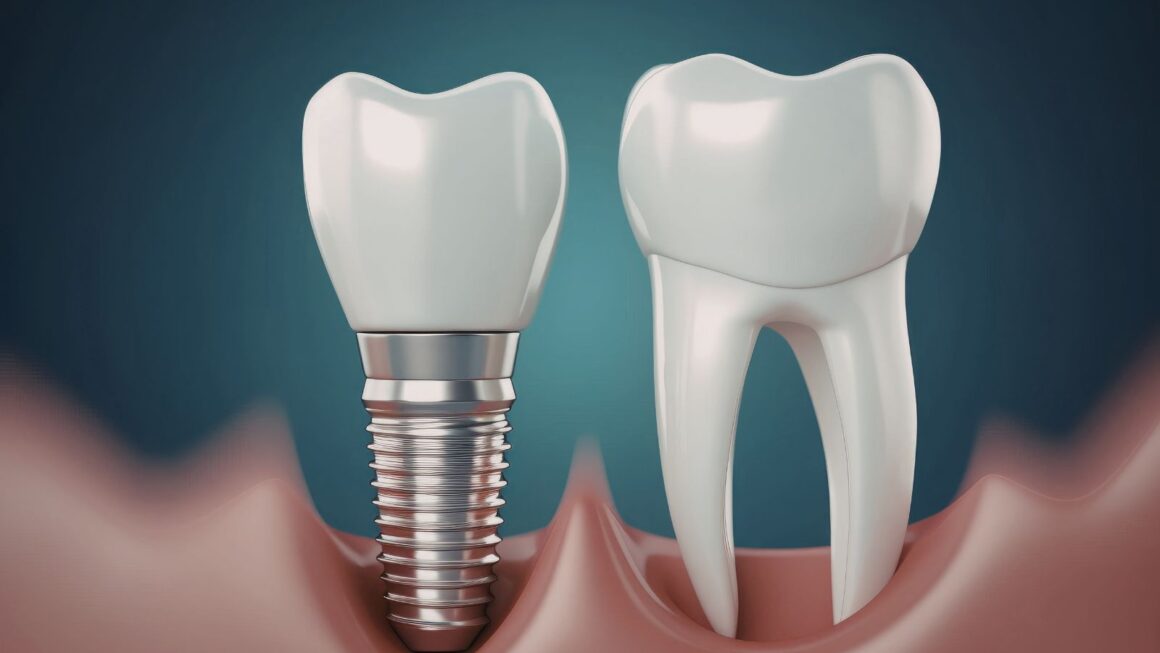
Osseointegration is the biological process where living bone grows onto and around an implanted material, creating a mechanically stable connection. In dentistry this usually involves a titanium or titanium-alloy post placed into your jawbone; bone cells (osteoblasts) deposit matrix onto the implant surface, and that matrix mineralizes to form rigid contact.
Successful osseointegration depends on implant surface properties, surgical technique, local bone quality, and your systemic health. Microscopic surface roughness and biocompatible coatings encourage faster bone apposition. Immediate mechanical stability at placement reduces micromotion that can disrupt bone formation.
Discovery by Dr. Per-Ingvar Brånemark
Dr. Per-Ingvar Brånemark first documented the phenomenon in the 1950s after observing that titanium chambers became firmly attached to rabbit bone. He translated that observation into controlled clinical experiments, demonstrating predictable long-term integration between titanium and human bone.
Brånemark introduced standardized implant designs, protocols for atraumatic surgical placement, and the concept of allowing a healing period before loading the implant. His work shifted implants from experimental to routine practice and established clinical criteria—primary stability, absence of infection, and appropriate healing time—for predictable outcomes.
Applications in Modern Dentistry
You encounter osseointegration most commonly with single-tooth implants, implant-supported bridges, and full-arch prostheses. Clinicians use it to replace one tooth or to anchor complete dentures, restoring chewing function and facial support.
Modern practice applies protocols that match implant type and surface to bone density and location. Examples include shorter healing times with roughened surfaces, guided bone regeneration when bone volume is insufficient, and immediate-loading in selected cases where initial stability is high. Patient factors—smoking, uncontrolled diabetes, and poor oral hygiene—remain critical determinants of success.
Biological Process of Osseointegration
Osseointegration depends on coordinated cellular responses, implant surface interactions, and mechanical conditions that together determine how bone forms and bonds to an implant. You’ll see how bone heals at the implant site, how surface chemistry and texture guide cells, the sequential stages of bone growth, and the key factors that influence early and long-term stability.
Bone-Healing Mechanism
When an implant is placed, the local blood clot forms immediately and brings platelets and signaling proteins to the site. Those signals recruit inflammatory cells and mesenchymal stem cells (MSCs) that differentiate into osteoblasts—the bone-forming cells you need for a stable implant.
Osteoblasts deposit a mineralized matrix directly onto the implant surface or onto newly formed woven bone nearby. Over weeks to months, remodeling replaces woven bone with stronger lamellar bone; osteoclasts resorb excess or poorly organized bone while osteoblasts rebuild, improving mechanical integration.
Control of micro-motion matters: small, controlled loading can stimulate bone formation, while excessive movement promotes fibrous tissue instead of bone, which reduces long-term fixation.
Role of Implant Surface Properties
The implant surface acts as the platform that cells interact with first. Surface chemistry (oxidation state, elemental composition) affects protein adsorption within seconds to minutes, and that adsorbed protein layer dictates which cell types attach and how they behave.
Surface topography at micro- and nano-scale alters cell orientation, adhesion strength, and differentiation. Roughened or porous titanium surfaces increase surface area for bone contact and enhance osteoblast activity compared with polished surfaces. Coatings such as calcium-phosphate or bioactive ceramics can further accelerate bone bonding by providing a chemical match to native mineral.
Sterility and absence of cytotoxic residues remain critical because contaminants impede cell attachment and can provoke chronic inflammation that undermines osseointegration.
Stages of Bone Growth
Primary stages start with hemostasis and inflammation in the first days. Platelets and immune cells release growth factors like PDGF and BMPs that recruit MSCs and endothelial cells, promoting initial vascularization.
The reparative phase follows: MSCs differentiate into osteoblasts and produce a collagen-rich osteoid that mineralizes into woven bone within 1–6 weeks depending on site and patient factors. New blood vessels form in parallel to support metabolic demands.
Remodeling is the longer-term stage where woven bone converts to lamellar bone and functional architecture aligns along load paths. This stage can continue for months and determines the final bone-to-implant contact percentage and mechanical strength.
Factors Affecting Implant Stability
Primary stability stems from mechanical engagement with cortical bone at placement; surgical technique and bone density determine this initial fixation. If you have low bone density or use overly aggressive drilling, you risk reduced primary stability.
Secondary stability arises from biological fixation through bone formation and remodeling. Systemic factors such as smoking, uncontrolled diabetes, and certain medications (e.g., bisphosphonates, corticosteroids) impair cellular function and slow osseointegration.
Local factors matter too: infection, excessive micro-motion, poor vascular supply, and inappropriate implant size or position all reduce bone formation at the interface. You can improve outcomes by optimizing implant macro-design, using appropriate surface treatments, and managing patient health to support predictable osseointegration.
Why Implants Fuse to the Jawbone
Osseointegration depends on three critical elements: the implant material and surface, the cellular and molecular reactions at the bone-implant interface, and the clinical management that preserves bone health over time.
Biocompatibility of Titanium and Other Materials
Titanium and titanium alloys interact favorably with bone because they form a stable, inert oxide layer on their surface. That oxide layer resists corrosion and reduces inflammatory responses, so your body recognizes the implant as compatible rather than foreign.
Surface treatments—such as sandblasting, acid etching, or coatings like hydroxyapatite—increase surface roughness and chemical activity. Those changes raise the implant’s surface area and provide micro- and nanoscale features where bone cells (osteoblasts) can attach and proliferate.
Material choice also affects mechanical properties. Titanium’s modulus of elasticity is closer to bone than many metals, which reduces stress shielding and encourages load transfer to surrounding bone. Modern alternatives, such as zirconia, offer similar biocompatibility with different esthetic and tissue-response profiles, but clinical evidence and handling characteristics vary by material.
Molecular Bonding at the Bone-Implant Interface
When you receive an implant, a sequence of molecular events begins within minutes to days. Blood proteins adsorb onto the implant surface first; their pattern determines which cells attach next. Proteins like fibronectin and vitronectin mediate cell adhesion, guiding mesenchymal stem cells and osteoprogenitors to the site.
Those cells differentiate into osteoblasts and secrete extracellular matrix—primarily collagen—which mineralizes into new bone. Over weeks to months, this new bone integrates tightly with the implant surface through physical interlocking and biochemical bonds involving calcium phosphate minerals.
Cell signaling pathways (e.g., BMPs, Wnt) modulate osteogenesis locally, while inflammatory cytokines influence remodeling. Controlled inflammation early on is necessary; excessive inflammation or infection disrupts protein adsorption and cell recruitment, undermining bonding and risking fibrous encapsulation instead of true osseointegration.
Maintenance of Long-Term Stability
Long-term stability hinges on mechanical loading, bone quality, and soft-tissue health around the implant. You maintain stability by ensuring functional loading distributes forces through the implant to the bone, which stimulates bone remodeling and preserves density. Immediate overloading or improper prosthetic fit can cause micro-movements that impede bone formation.
Peri-implant bone loss stems from factors you can control: poor oral hygiene, smoking, untreated periodontal disease, and systemic conditions like uncontrolled diabetes. Regular professional evaluations, radiographic monitoring, and routine hygiene reduce risk. If bone loss begins, interventions such as debridement, soft-tissue management, or bone grafting can re-establish a stable environment for osseointegration to persist.
Advancements and Challenges in Osseointegration
Recent improvements focus on better implant surfaces, surgical precision, and integrated prosthetic interfaces. You’ll see faster bone integration, enhanced sensory feedback, and clearer strategies to reduce infection risk.
Technological Innovations
Manufacturers now use roughened and porous titanium surfaces to increase bone contact and mechanical interlock. These surface topographies and coatings (including hydroxyapatite and bioactive peptides) promote osteoblast attachment and faster bone ingrowth.
Press-fit implant designs and custom 3D-printed fixtures let surgeons match implant geometry to your bone, improving primary stability and reducing micromotion during early healing. Navigation systems and patient-specific drilling guides increase placement accuracy, lowering the chance of implant failure from malposition.
Researchers also integrate electrical stimulation and drug-eluting coatings to accelerate bone formation and limit bacterial colonization. On the prosthetic side, direct skeletal attachment systems now support improved load transfer and, in some cases, better sensory feedback through osseoperception, helping you sense pressure and position more naturally.
Potential Complications
The main risks remain infection at the skin-implant interface, implant loosening from insufficient bone contact, and periprosthetic fractures. Skin breaches create a persistent pathway for bacteria; management requires meticulous soft-tissue handling, regular hygiene protocols, and sometimes long-term antibiotics.
Poor primary stability or excessive micromotion can prevent bone from bonding to the implant, leading to fibrous tissue formation and eventual loosening. Smoking, uncontrolled diabetes, and poor bone quality (osteoporosis) increase that risk.
Surgeons mitigate complications with preoperative bone assessment (CT or DEXA), staged loading protocols, and careful patient selection. When problems arise, revision surgery or adjunctive bone-grafting and anti-infective strategies may be necessary to restore a stable implant environment.