
Bone loss can come from infection or from losing a tooth, and the cause changes what you should do next. Gum disease erodes the bone that anchors teeth through chronic infection and inflammation, often progressing slowly and threatening multiple teeth; losing a tooth causes localized bone resorption where the socket no longer receives stimulation. For those considering dental implant surgery in Inglewood, CA, knowing whether infection or lack of stimulation is driving your bone loss is critical, because the treatment and prevention strategies differ significantly.
You’ll learn how clinicians diagnose each type, what signs to watch for, and which treatments — gum therapy, bone grafting, or implant planning — match each situation. That clarity helps you protect surrounding teeth, plan for replacements, and avoid further jawbone loss.
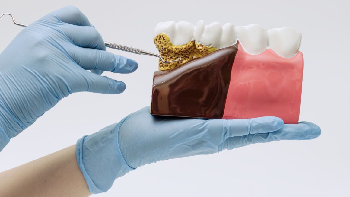
Understanding Bone Loss from Gum Disease
Gum disease destroys the structures that hold your teeth in place: the periodontal ligament and the surrounding alveolar bone. You will learn what causes this bone loss, how it progresses over time, and how clinicians diagnose it.
Causes of Bone Loss in Periodontal Disease
Periodontal bone loss starts with bacterial plaque forming a biofilm on tooth surfaces. Your immune response to that biofilm creates inflammation; inflammatory mediators (cytokines, prostaglandins) stimulate cells that break down bone (osteoclasts).
Risk factors speed the process. Smoking reduces blood flow and impairs healing, diabetes elevates inflammatory responses and impairs immune control, and poor oral hygiene allows plaque to mature into calculus that sustains infection. Certain medications and genetic predisposition can also increase your susceptibility.
Local factors like crowded teeth, overhanging restorations, or deep gum pockets concentrate plaque and make mechanical cleaning difficult. Addressing both the bacterial load and the host/environmental risks matters to slow or stop bone destruction.
Progression and Long-Term Effects
Bone loss often begins as localized resorption around a tooth root and can progress to generalized bone height reduction across multiple teeth. Early stages may show mild vertical defects or shallow pockets; advanced disease creates wide bone loss, furcation involvement on molars, and mobility.
As bone support diminishes, your teeth can shift, become sensitive, and eventually loosen or fall out. Altered bite mechanics from lost teeth may cause additional wear and jaw discomfort. Untreated severe periodontitis increases the need for complex restorations, extractions, or prosthetic replacements such as implants or dentures.
Diagnosis of Bone Loss Linked to Gum Disease
Clinicians combine clinical exam and imaging to diagnose periodontal bone loss. Your dentist measures pocket depths, checks for bleeding on probing, furcation involvement, and tooth mobility. Probing patterns that deepen over time point to active disease.
Radiographs (bitewing and periapical, sometimes CBCT) reveal bone height, patterns of loss (vertical vs. horizontal), and localized defects. Labs or medical history review identify systemic contributors like uncontrolled diabetes or medications affecting bone metabolism. Treatment planning depends on severity, tooth prognosis, and your overall health.
Bone Loss After Tooth Extraction
After a tooth is removed, the jawbone that once supported its root loses the mechanical stimulation it needs to maintain volume. That loss of stimulation, along with local remodeling and healing processes, drives predictable patterns of bone resorption that affect ridge height and width.
Mechanisms of Post-Extraction Bone Resorption
When you lose a tooth, the periodontal ligament and its function disappear. The ligament previously transmitted chewing forces to the alveolar bone; without that stimulus, osteoclast activity increases and osteoblast-mediated bone formation declines, causing net bone resorption.
Soft-tissue and inflammatory responses also influence remodeling. Clot formation and early socket healing recruit cells that reshape the socket; if infection or chronic inflammation occurs, resorption accelerates. The thin facial (buccal) plate—especially in the anterior maxilla—is prone to rapid collapse because it is often <1–2 mm thick.
Local anatomy and systemic factors modify the response. Smoking, uncontrolled diabetes, and certain medications (e.g., bisphosphonates, steroids) increase resorption risk. Immediate grafting or implant placement can preserve ridge volume by maintaining space and providing scaffold or load stimulation.
Timeline and Patterns of Bone Loss
Bone changes begin within days and continue for months to years. You can expect most dimensional change in the first 3–6 months after extraction, with notable horizontal (width) loss often exceeding vertical loss.
Typical pattern:
- First week: clot organization and early soft-tissue closure.
- 1–3 months: peak bone remodeling; substantial ridge contraction.
- 3–12 months: ongoing remodeling with slower volume reduction; stabilization often by 6–12 months.
Anterior sites and areas with thin cortical plates show greater early collapse. Posterior mandibular sockets may retain more height but still lose width. If you plan prosthetic replacement, aim for ridge preservation at the time of extraction or implant placement within the early healing window to limit dimensional changes.
Clinical Indicators and Assessment
Evaluate sockets clinically and with imaging to quantify loss. On exam, you’ll note depression of the ridge, reduced buccolingual width, and changes to soft-tissue contour that complicate prosthetic emergence profiles.
Use periapical or panoramic radiographs for vertical height trends; cone-beam CT gives precise 3D measurements of width and height. Key measurements include:
- Buccolingual ridge width at the crest and 1–5 mm below crest.
- Vertical ridge height relative to adjacent teeth or anatomical landmarks.
Monitor for signs of infection, delayed healing, or bone sequestra. Document baseline measurements at extraction and reassess at 3 and 6 months to guide grafting, implant timing, or prosthetic planning.
Key Differences Between Bone Loss from Gum Disease and Tooth Extraction
You should know how cause, tissue involvement, and treatment options differ because they determine diagnosis, timing of intervention, and restoration choices.
Underlying Biological Processes
Gum disease (periodontitis) causes bone loss through a chronic bacterial infection. Bacterial plaque triggers inflammation that activates immune cells and enzymes which break down the periodontal ligament and alveolar bone supporting your teeth. This process is gradual and can be localized around one tooth or spread across multiple sites; X-rays often show horizontal or vertical bone defects and loss of the bone crest.
Bone loss after tooth extraction is primarily a mechanical and physiological remodeling response. When a tooth and its periodontal ligament are removed, the alveolar ridge loses the mechanical stimulus that maintains bone volume. Resorption usually happens fastest in the first 3–6 months and affects the socket width and height, with more pronounced loss on the buccal (cheek) side.
Impact on Surrounding Oral Structures
With periodontitis, adjacent teeth and soft tissues suffer progressive attachment loss. You may see gum recession, increased tooth mobility, pocket formation, and migrating teeth. Infection-driven bone defects can undermine multiple neighboring roots, making teeth non-restorable without periodontal surgery or extraction.
After extraction, the defect is confined initially to the socket and adjacent ridge. Surrounding teeth remain structurally intact unless they were compromised by preexisting disease. However, ridge collapse alters prosthetic space and adjacent tooth alignment over time. If you don’t place an implant or perform socket preservation, you’ll typically lose horizontal width and some vertical height, complicating future restorations.
Implications for Dental Treatment and Restoration
Treating bone loss from gum disease focuses on infection control and regenerating lost support. You’ll likely receive scaling and root planing, systemic or local antimicrobials, and possibly flap surgery with bone grafting or guided tissue regeneration (GTR) to rebuild defects. Long-term maintenance and improved oral hygiene are essential to prevent recurrence.
After extraction, treatment emphasizes ridge preservation and prosthetic planning. Options include socket grafting at the time of extraction, immediate or delayed implant placement, or planning for removable/fixed prostheses with possible ridge augmentation. The timing of grafting and implant placement depends on soft-tissue healing and whether infection is present; prompt intervention preserves bone and simplifies later restoration.
Prevention and Management Strategies
You can slow or stop bone loss by controlling infection, replacing missing teeth promptly, and improving oral and overall health. Specific actions include professional periodontal care, timely restorative procedures, and targeted lifestyle changes.
Dental Care to Minimize Bone Loss
Keep plaque and gum inflammation under control with twice-daily brushing using a soft brush and interdental cleaning once a day. Use a fluoride toothpaste and consider an antimicrobial mouthwash if your dentist recommends it after assessment.
Schedule professional cleanings every 3–6 months if you have periodontitis; clinicians will perform scaling and root planing to remove subgingival biofilm and calculus that drive bone destruction. Your dentist may prescribe localized antibiotics or host-modulation therapy when inflammation persists despite mechanical therapy.
Maintain regular radiographic monitoring (bitewings or periapicals) to track bone levels. If you notice receding gums, loose teeth, or shifting bite, contact your dentist promptly for evaluation.
Restorative Options for Lost Bone
When bone loss follows extraction or periodontal disease, discuss bone grafting to rebuild ridge volume. Options include autografts, allografts, xenografts, and synthetic grafts; your surgeon will recommend the material based on defect size, healing potential, and future restorative plans.
Guided bone regeneration (GBR) uses barrier membranes to protect grafts and encourage new bone formation. If you plan implants, socket preservation at the time of extraction reduces ridge collapse and improves implant positioning.
For advanced defects, combination approaches—bone graft plus soft-tissue grafting—improve both function and esthetics. Your clinician will outline timelines: graft integration commonly takes 3–6 months before implant placement or definitive prosthetics.
Lifestyle Modifications and Professional Guidance
Quit smoking and limit alcohol; both impair bone healing and increase risk of periodontal progression. Improve nutrition with adequate calcium and vitamin D, and discuss supplementation with your physician if lab tests or diet suggest insufficiency.
Control systemic conditions such as diabetes by keeping HbA1c in target range, since hyperglycemia worsens periodontal inflammation and bone loss. Inform your dentist about osteoporosis medications (bisphosphonates, denosumab) because they affect surgical planning and healing.
Keep a treatment plan and maintenance schedule with your dental team. Follow-up visits for periodontal maintenance, hygiene reinforcement, and radiographic checks let your clinician catch relapse early and adjust therapy as needed.